
This article presents a clinical case of successful treatment of a 24-year-old female patient diagnosed with severe generalized periodontitis. The distinctive feature of this case is the achievement of complete bone tissue regeneration, including the formation of an interdental bone peak that extended 2 mm above the original level of the buccal cortical plate. The result was obtained using the osteoplastic material Bio-Oss without the application of barrier membranes or enamel matrix proteins. Six months after surgery, elimination of tooth mobility, periodontal pockets, and inflammatory manifestations was documented.
In 2023, a 24-year-old patient (Olga) presented to the Periodontal Center Periostom with a diagnosis of severe periodontitis. At the initial consultation, generalized inflammation of the periodontal tissues, pronounced tooth mobility (Miller Class I and II), probing depths of 6 to 12 mm, recurrent periodontal abscesses, and bone loss reaching 50% of the original level were observed. Due to the severity of the clinical presentation, tooth extraction of most teeth had been recommended at other dental facilities.
The treatment plan included surgical interventions aimed at eliminating intraosseous defects. In the area of greatest interest for this observation, the initial depth of the intraosseous defect measured relative to the buccal cortical wall was 3 mm.
During the procedure, debridement of the intraosseous defects was performed, and the defects were filled with granules of bovine-derived osteoplastic material (Bio-Oss). Barrier membranes and enamel matrix proteins (EMD) were not used. Regeneration was achieved through the potential of the patient’s own tissues and the osteoconductive properties of the material.
Six months after surgery, follow-up diagnostics were performed. Visualization was carried out using cone‑beam computed tomography (CBCT), as well as intraoperatively due to the need to address periodontal pockets in an adjacent segment. The evaluation revealed:
1. Clinical parameters: tooth mobility was absent; probing depths were undetectable; bleeding and suppuration were absent; periodontal abscesses and halitosis did not recur.
2. Radiological and intraoperative findings: complete restoration of bone tissue was documented. In the area of the previously existing intraosseous defect, vertical bone growth exceeding the original level of the buccal wall was observed. The initial defect depth (3 mm relative to the buccal wall) was eliminated, and an interdental bone peak had formed, extending 2 mm above the buccal wall. The total volume of newly formed, high‑quality bone in this area amounted to 5 mm.
The presented case demonstrates the possibility of achieving vertical bone regeneration (formation of a bone peak above the level of the buccal wall) in the oral cavity without the use of membrane techniques or growth factors.
The uniqueness of this observation also lies in the ability to verify the result intraoperatively after six months. Typically, assessment of the quality and volume of newly formed tissue without invasive intervention is challenging. In this case, addressing intraosseous defects in the adjacent segment allowed for visual confirmation not only of the quantitative bone gain (5 mm total) but also of its high quality, which is suitable for long‑term stability of the dentition.
The surgical phase was performed by the Chief Physician of the PerioStom Periodontal Center, surgeon‑periodontist, implantologist Grigory Sergeyevich Isayan.
On April 5, 2025, in Moscow, I presented this clinical case to Professor Dr. Pierpaolo Cortellini and wish to express my immense gratitude once again for the knowledge and techniques he has imparted through international training, which help us save thousands of teeth for our patients, including this young woman who, at the age of 24, had been advised to have most of her teeth extracted.
P.S.: The working title for this clinical case is Dr. Isayan’s Peak
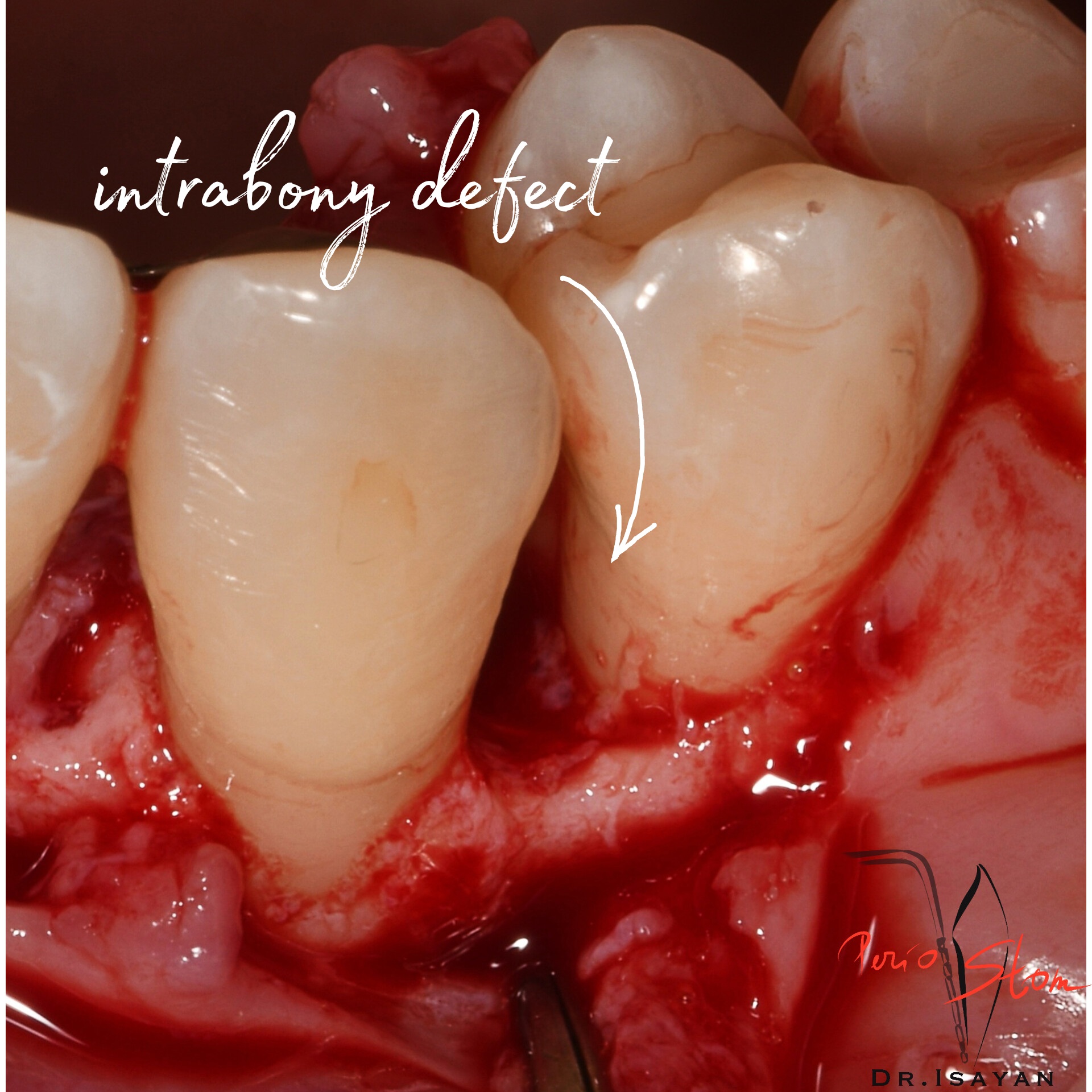
intrabony defect
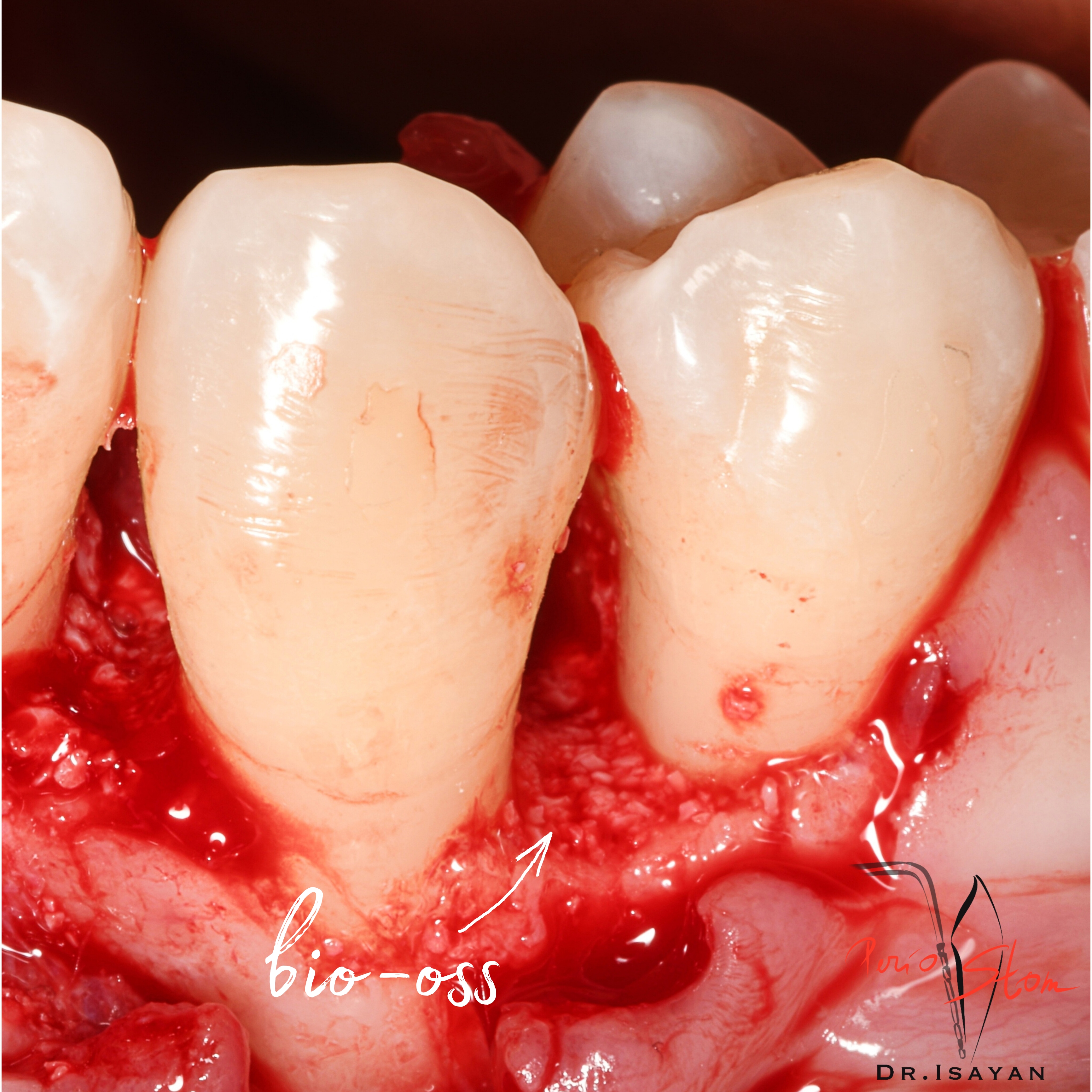
bio-oss-L
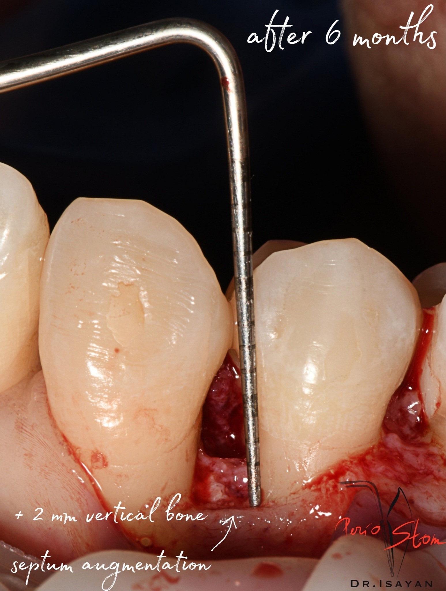
+2 mm vertical bone septum augmentation
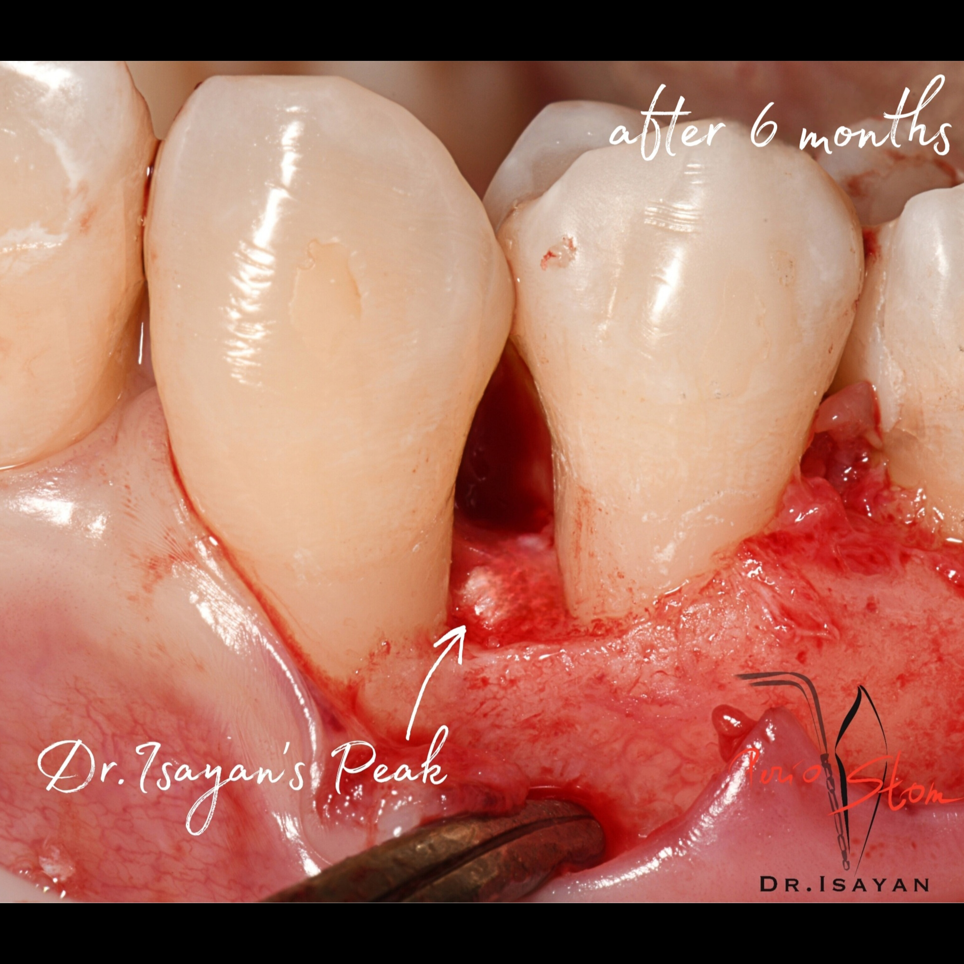
after 6 months, dr.Isayans Peak
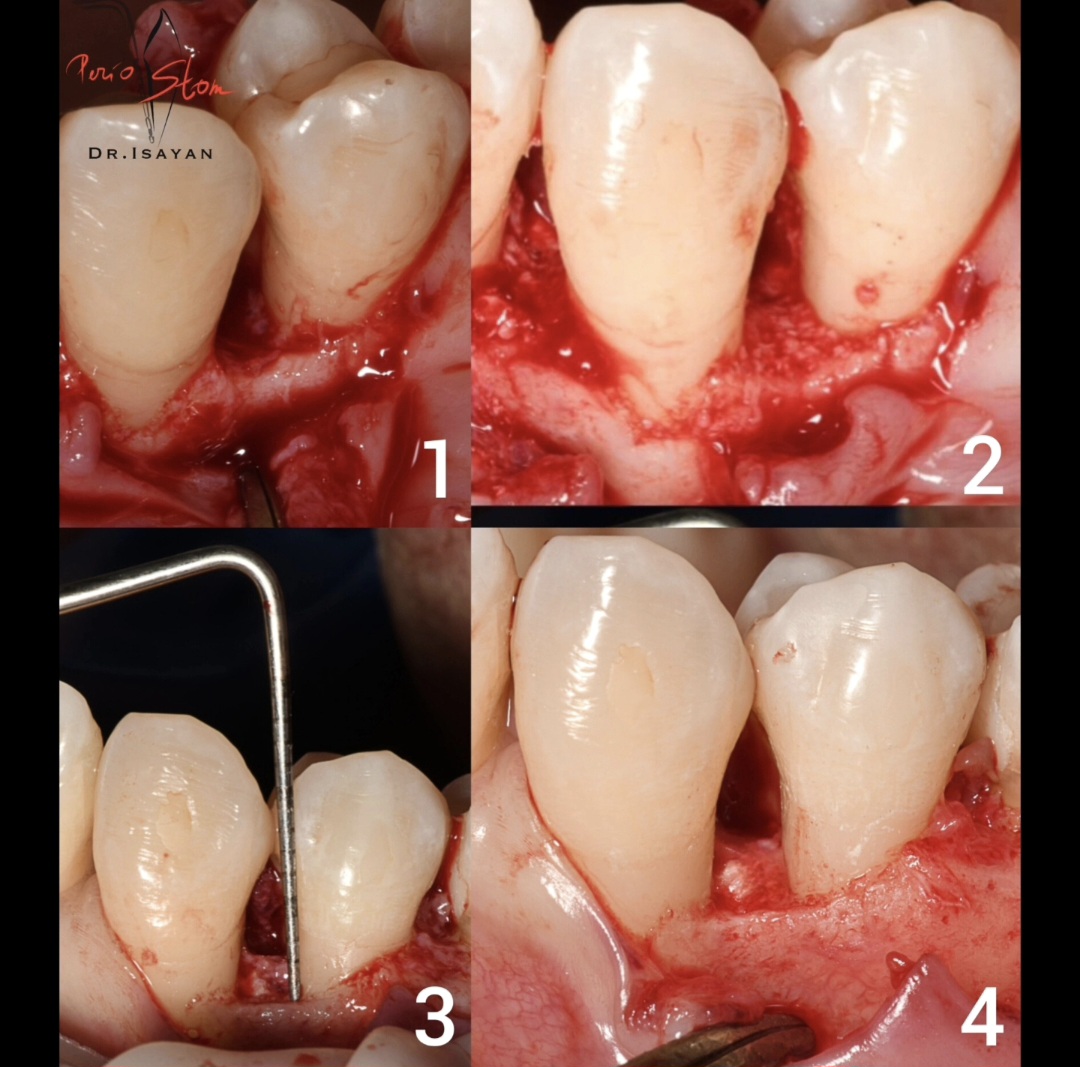
stage by stage
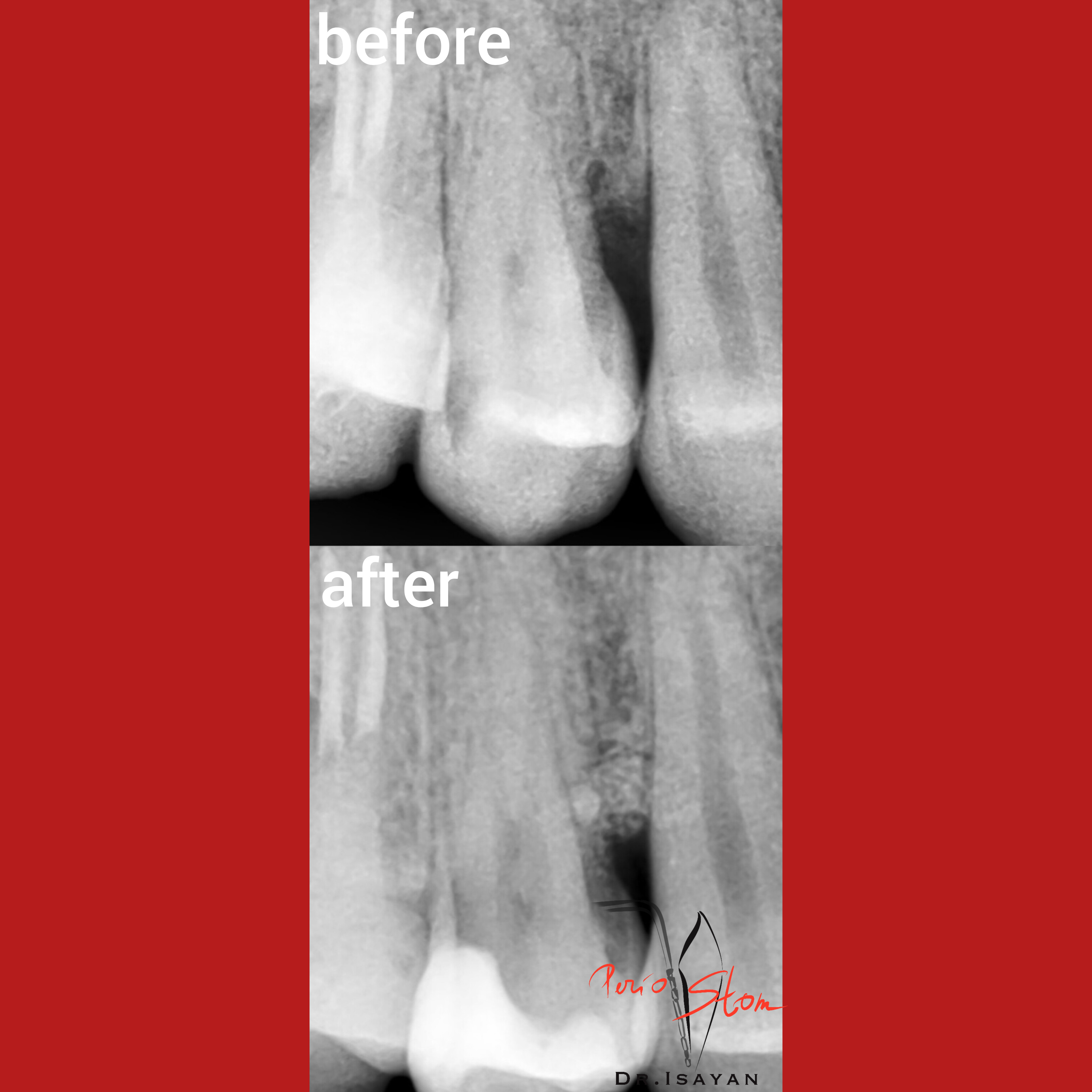
x-ray
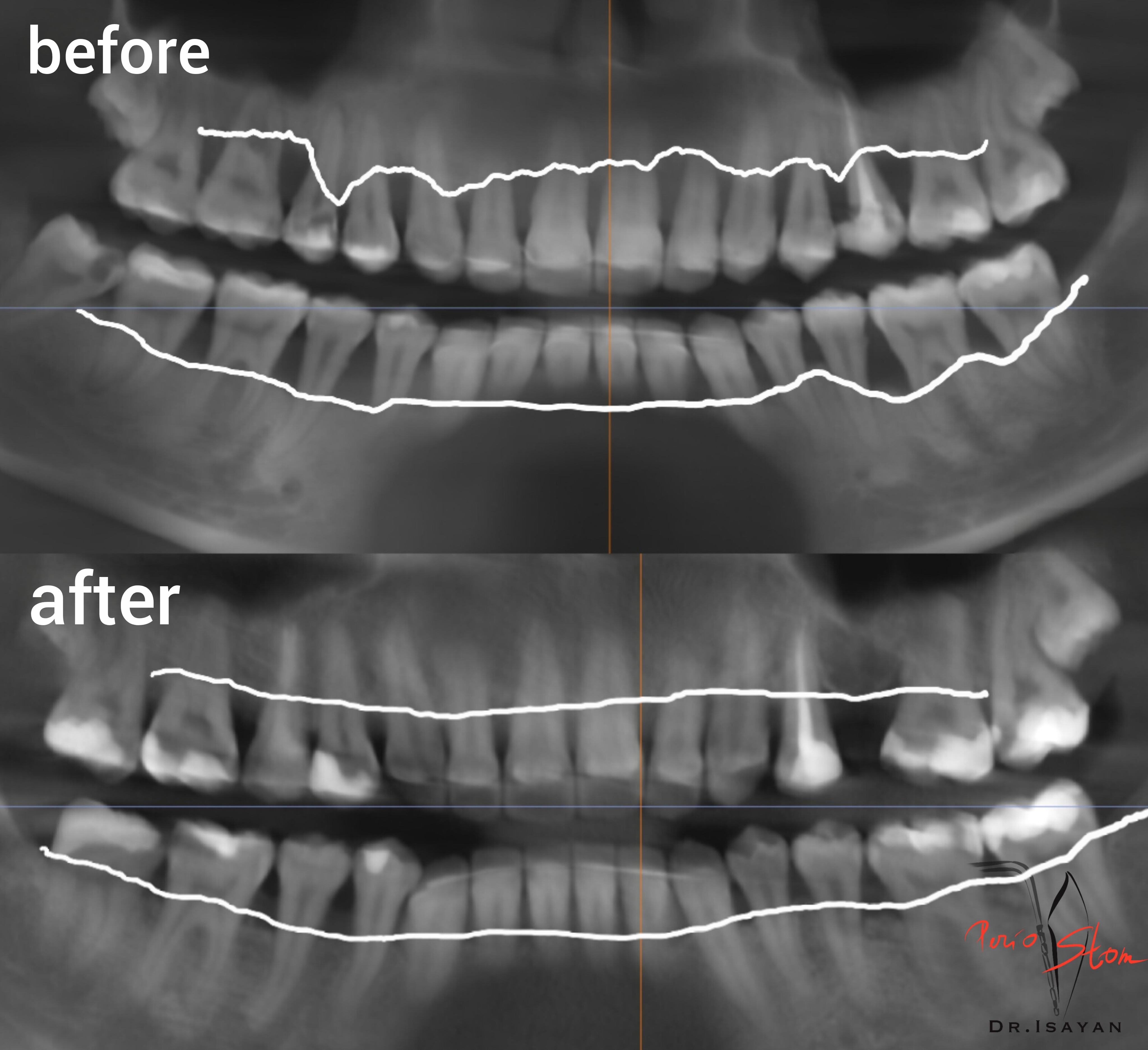
panoramic dental x-ray
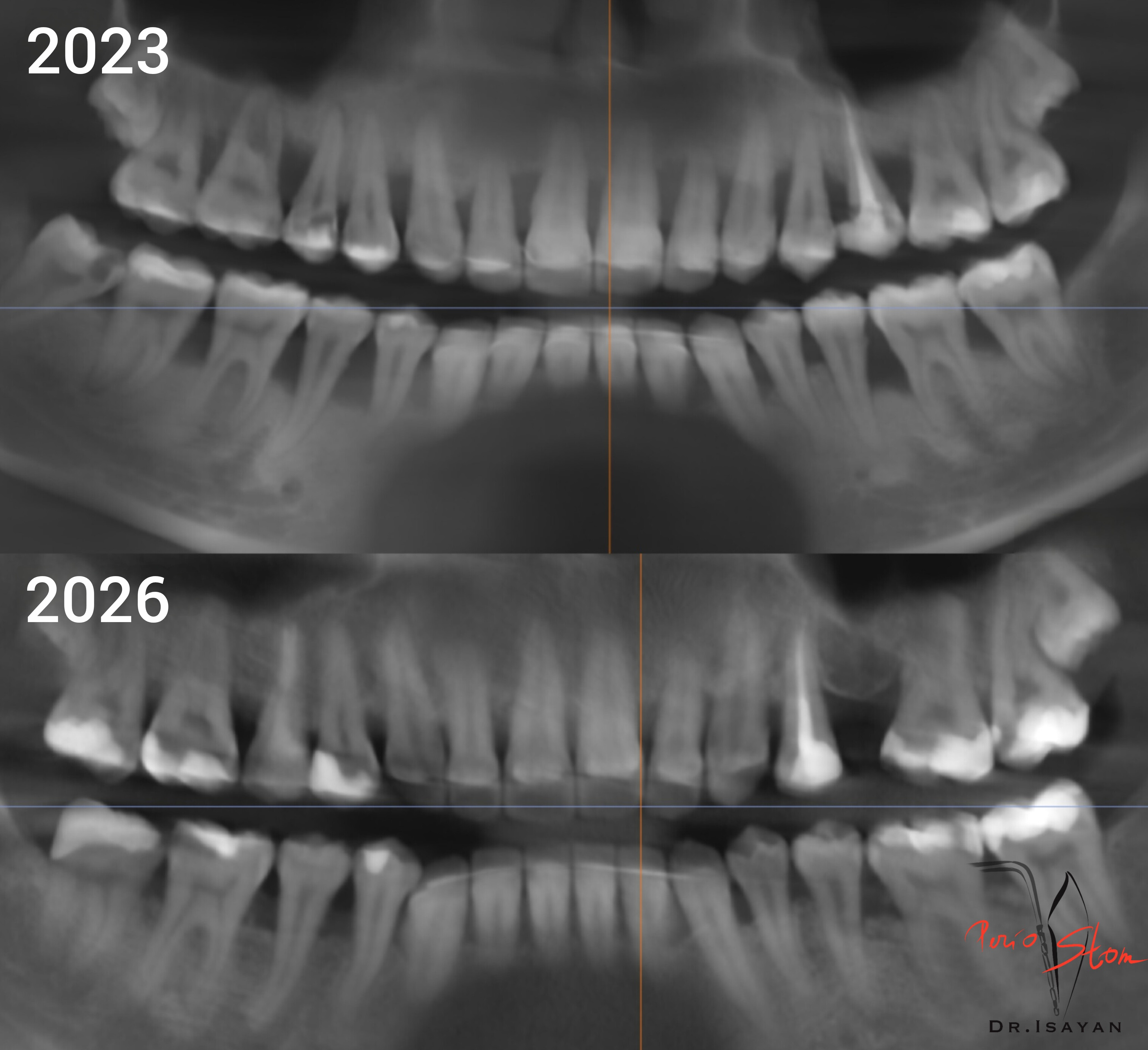
panoramic dental x-ray, 2023-2026
Powered by